Country
ESP
Affiliation
Universidad Autónoma de Madrid

Despite decades of advances in magnetic imaging, obtaining direct, quantitative information with high spatial resolution remains an outstanding challenge. The imaging technique most widely used for local characterization of magnetic nanostructures is the Magnetic Force Microscope (MFM), which is indeed a very active topic of investigation [1]. Advantages of MFM include relatively high spatial resolution, simplicity in operation as well as sample preparation, and the capability to applied in situ magnetic fields to study magnetization process [2]. The tip engineering, the quantitative measurements, the correct interpretation of the resulting MFM images, the possibility of operate in different environments including liquid media to investigate biological samples [3], or the analysis of the loss of energy [4] are subjects of ongoing research that will be reviewed in this talk.
In particular, we will try to approach some of the challenges of MFM by following different routes. One route is the development of high-performance MFM probes with sub-10 nm (sub-25 nm) topographic (magnetic) lateral resolution by following different easy and quick low-cost approaches [5]. This allows one to not only customize the tip stray field, avoiding tip-induced changes in the sample magnetization, but also to optimize MFM imaging in vacuum (or liquid media [6]) by choosing tips mounted on hard (or soft) cantilevers, a technology that is currently not available on the market. On the other hand, the use of advanced MFM operation modes such as the combination of multifrequency modes [7] with the information obtained from the experimental dissipation of energy associated to tip-sample interactions [8] will be explored in order to improve the MFM capabilities.
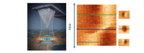
Figure1: Nonstandard MFM image of a Py nanodot (height ~30 nm). With this method, we can study the magnetization process of a single particle.
[1] O. Kazakova,et al. Journal of Applied Physics 125, 060901 (2019).
[2] E. Berganza, M. Jaafar, et al. Nanoscale, ,12, 18646-18653 (2020).
[3] M. Jaafar et al. ACS Appl. Mater. Interfaces, 6, 20936 (2014)
[5] M.Jaafar et al, Nanoscale, 12, 10090 –1009 (2020).
[6] P. Ares, M. Jaafar et al. Small 11,36, 4731-4736 (2015).
[7] VG Gisbert et al. Nanoscale 13 (3), 2026-2033 (2021).
[8] M. Jaafar and A. Asenjo, Appl. Sci., 11(22), 10507 (2021).